鲜花( 87)  鸡蛋( 1)
|
 - Small Wonders: Finalists From the Nikon Small World Competition

8 r- m8 l7 X: \9 }0 hHere we present ten of the finalists from Nikon’s 35th Annual Small World Photomicrography Competition, which recognizes photographs shot through a microscope. Contest winners will be announced on October 8. Until October 2, the public can select their favorites in the “Popular Vote” section of the Nikon Small World web site.% W$ D/ r4 B3 [: _2 l K2 o
Above: © Shamuel Silberman, Ramat-Gan, Israel+ h X! N0 n8 f! b3 |( s; b
Embryo of guppy fish (40X)' T E" v7 \* X: C" p; e/ s
Reflected light by fiber-optics5 {. t) v- R6 J: O( \
+ d' }6 X( P( C8 j% h3 W
- h. [0 b1 e4 B4 y9 V% u! W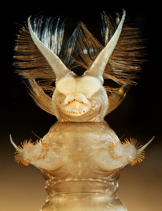
0 e8 a2 d4 e/ K© Fabrice Parais, DIREN Basse-Normandie, Hérouville-Saint-Clair, France7 @. c' G6 g% q P5 s, `
Atherix ibis (fly) aquatic larva (25x)
& _% F" ~- M9 E& R6 E1 k" WStereomicroscopy
' Q- W( l7 f4 ~) B0 [8 c+ e/ J ( w E2 [1 Y2 y% E/ z ( w E2 [1 Y2 y% E/ z
© Karie Holtermann, Rancho Cucamonga, California, United States8 a6 M/ R# T% v& v. ~# ?% V
Raindrop on butterfly wing (20X)8 J5 A% z3 _1 Z1 N
Differential interference contrast) S( g0 L1 j z) T1 a" l: \% Z
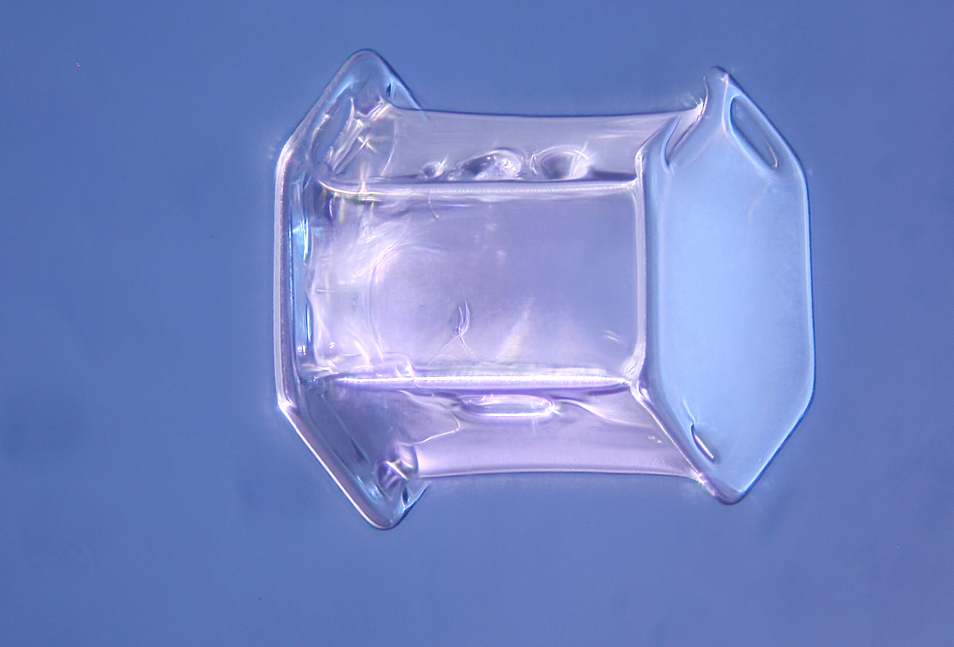 1 C! P" D$ o, c% I/ w 1 C! P" D$ o, c% I/ w
© Yanping Wang, Beijing Planetarium, Beijing, China4 q$ T) ?: i& K. ]/ W- u# z, a
Snowflake (40X)* M& r# |& p' l" R/ b% I9 v
Reflected and Transmitted Light
5 R0 B. B* f+ \: o, x
0 s7 x8 {( i( f2 g9 M" D/ a© Gerd A. Guenther, Düsseldorf, Germany4 i9 I# d( S# n0 t
Sonchus asper (spiny sowthistle) flower stem section (150X)3 q) A& l* O% Y+ t* Y) K) s
Darkfield/ B0 y% ^; M: o7 k7 X

% \6 b8 D* w s+ \© Norm Barker, Department of Pathology, Johns Hopkins University, School of Medicine, Baltimore, Maryland, United States
/ r) w' f- I6 e4 ?7 f# v3 B* PDinosaur bone, Jurassic period (15X)/ }; I+ S$ T q7 O9 P. e
Reflected light from fiber optic/ O, C( ?8 o; `

2 c% E7 w3 i( c1 N5 k+ J3 K© Viktor Sykora, Institute of Pathophysiology, First Medical Faculty, Charles University, Prague, Czech Republic
: G8 X6 V5 b# w4 A7 N; |) mHoya carnosa (wax plant) flower (10x)
& ^* t5 h6 {; c' t$ B H( w, L7 mDarkfield
# O1 Y2 b$ [* c0 ]1 W3 n4 l2 S
' I8 U3 a+ V8 S© Daniel Vega, Madrid, Spain$ d( Q) ~; s+ i+ Q, @. i% d4 d1 N
Gall (plant tissue growth) formed by Trigonaspis mendesi (4X)" C* P/ y Q" Y% |* O/ q! M7 v
Incident light and transillumination/ E. E8 C# u3 Y" V

6 S% u8 m9 ^9 m5 ~+ F# h/ q9 d! A2 g© Massimo Brizzi, Microcosmo Italia, Empoli, Firenze, Italy
% M- Y. m6 N& Q% ]6 ?Snail eggs (200x)7 P, r0 T1 c# k' `6 H, W
Differential Interference Contrast: T# f) q1 J; T8 | F: x/ z4 B0 ~

( p9 u \& d/ ^3 A- k© Frederique Ruf-Zamojski, California Institute of Technology, Pasadena, California, United States
% h M" @$ M0 k1 e* b& a) H( nZebrafish embryo, 22 hours post-fertilization, living specimen (40X)
3 c, g0 b `8 y% ^8 QConfocal
& j, k. W) q9 M9 qTags: Photomicrography
|
|

















 狗仔卡
狗仔卡
 发表于 2010-10-25 02:35
 提升卡
提升卡 置顶卡
置顶卡 沉默卡
沉默卡 喧嚣卡
喧嚣卡 变色卡
变色卡 显身卡
显身卡


 发表于 2010-10-25 09:38

 油+水?支持你为GF1上微距。
 都是好片。
都是好片。


 发表于 2010-10-26 14:48